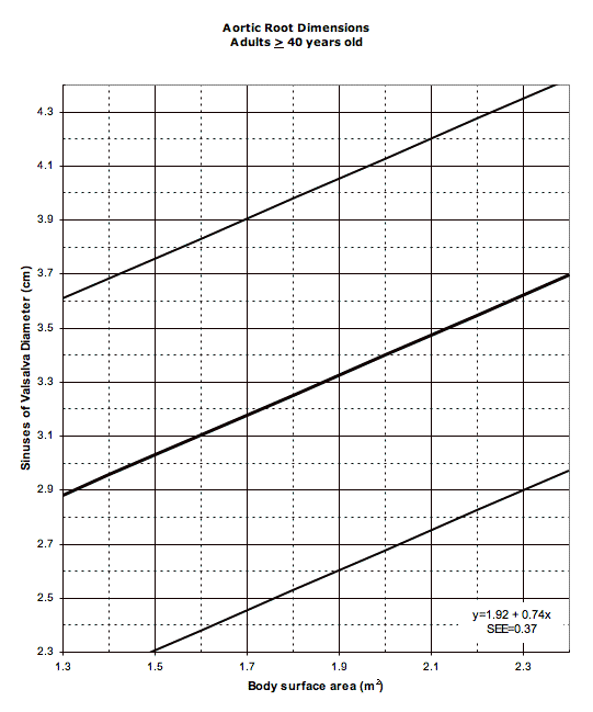
Marfan Syndrome Aortic Root Dimensions
Establishing whether the aortic root is
enlarged requires three things.
The first is
what the "normal" aortic root dimension is,
the second is
how much above this should be considered enlarged,
and the third is an accurate measurement of the
patient's aortic root size.
The reference for the "normal" root dimension requires data and
proper application or use of that data. These data need to
include important factors that may limit the use of the refence
such as gender, race, or others that might systematically
affect the results.
How much above the threshold is used for defining enlargement
is arbitrary but contributes to the sensitivity and specificity
of this as a diagnostic criterion.
This definition is independent of the degree of enlargement
that needs medical intervention.
The aortic root measurement for the patient needs to be
carefully done using a technique that matches the technique
used to generate the reference value.
The diagnostic criteria described by DePaepe and colleagues
includes several nomograms for evaluating aortic root dimensions
that were reproduced from a publication by Dr. Roman and colleagues1.
These normalize the aortic dimension to the body surface area.
The aortic dimension is measured at the sinuses of Valsalva
by cross-sectional 2-dimensional surface echocardiography
from the parasternal long-axis view.
No description of the method to calculate
the body surface area is given, so for the Automated Marfan
Syndrome Checklist the Mosteller2 formula is used:
BSA(m2) = ([Height(cm) x Weight(kg)] / 3600)1/2
The nomograms define the 95% confidence intervals.
This is +1.96 times the standard error (SEE or standard error of
the estimate on the graphs).
Values above the top end of this interval are considered "enlarged."
A re-creation of these nomograms are reproduced below for
easy reference.
The publication by Roman and
colleagues1 states that the infant and child nomogram was
based on data from
from 52 infants and children from ages 1 month to 15 years
and adult nomograms were based on data from 135 adults
from 20 to 74 years of age.
This leaves patients in the 15 to 20 year old range as not
being represented in either curve. For the calculations
used for the Automated Marfan Syndrome Checklist ages under 17.5 years
are considered infants or children and 17.5 years and older
are considered as adults. Additionally, no description of race
is included so these same nomograms are used for all individuals.


In each of these graphs the central diagonal line represents the
expected aortic root diameter and is flanked by lines representing
the 95% confidence intervals.
Clicking on the graphs will display a larger version.
More recently Dr. Pettersen and colleagues published data from a much
larger cohort of infants to adolescents and a number of echocardiographic
measurements3.
These data are from 782 patients age 1 day to 18 years, a little better
than the 52 from.
The Pettersen3 data use the Dubois body surface area
formula for normalization (example
BSA calculator
page).
BSA(m2) =
0.20247 x ([Height (cm)] / 100)0.725 x [Weight (kg)]0.425
The aortic root measurements are made at the sinuses of valsalva hinge
points from the parasternal long axis view at their maximum systolic
dimention.
The article provides a table with coefficients and a formula for
calculating Z-scores. The coefficients for the sinuses of Valsalva
measurements are -0.5 for the intercept (β0),
2.537 for BSA (β1),
-1.707 for BSA2 (β2),
0.420 for BSA3 (β3),
and 0.012 for the mean square error (MSE),
with an R2 of 0.916.
References:
1. Roman MJ, Devereux RB, Kramer-Fox R, O'Loughlin J: Two-dimensional echocardiographic arotic root dimensions in normal children and adults. Am J Cardiol 1989 Sept 1;64:507-512
2. Mosteller RD: Simplified Calculation of Body Surface Area. N Engl J Med 1987 Oct 22;317(17):1098 (letter)
3. Pettersen MD, Wei Du, Skeens ME, Humes RA: Regression Equations for Calculation of Z Scores of Cardiac Structures in a Large Cohort of Healthy Infants, Children, and Adolescents: An Echocardiographic Study. J Am Soc Echocardiogr. 2008 Aug;21(8):922-34
This description is a part of the
Automated Marfan Syndrome Checklist.


